 化工儀器網
化工儀器網技術文章
選購心肌細胞成熟測定裝置,心肌成熟試驗裝置
閱讀:476 發布時間:2020-7-13
心肌成熟試驗裝置-功能性iPSC誘導的心肌細胞纖維
Cardiomyocyte maturation assay
Functional iPSC induced cardiomyocyte fibers
心肌成熟試驗
功能性iPSC誘導的心肌細胞纖維
讀出
跳動頻率,肉瘤觀察
標準文化限制
在標準培養物中,源自多能干細胞的心肌細胞具有隨機形狀,沒有組織,因此顯示出不成熟的特性。
心肌細胞成熟測定
當在帶有線性黏附提示的底物中培養時,心肌細胞會獲得細長的形狀,從而調節細胞的收縮力,并建立并維持肌原纖維的排列方式,并改善并規則地打動細胞。
例
心肌細胞以線型組織[1],以及肌原纖維排列在收縮力中的作用。
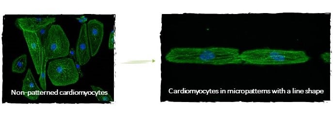
微模式下成熟的心肌細胞
參考資料
[1] hPSC-CM無圖案(左圖)和在線微圖案(右圖)通過熒光顯微鏡成像。 Dussaud S.,Jouve C.和Hulot J.S.,2018年。這些圖像是由Hulot教授(HôpitalEuropéenGeorges-Pompidou)及其團隊使用4Dcell產品提供的。
[2] Max R. Salick,Wendy C. Crone。等。在人類胚胎干細胞衍生的心肌細胞,生物材料中的微模式寬度依賴的Sarcomere發展。 2014年5月。 35(15):4454-4464。
READ-OUTS
Beating frequency, observation of sarcomeres
STANDARD CULTURE LIMITATION
In standard cultures, cardiomyocytes derived from pluripotent stem cells have random shapes, are not organized thus showing immature properties.
CARDIOMYOCYTE MATURATION ASSAY
When cultured in a substrate with line adhesive cues, cardiomyocytes acquire elongated shapes, which regulates cell contractility and establishes and maintains myofibril alignment and an improved and regular cell beating.
EXAMPLE
Organization of cardiomyocytes in line patterns [1], and effect of myofibril alignment in contractility forces.
REFERENCES
[1] hPSC-CM non patterned (left image) and on line micropatterns (right image) imaged by fluorescence microscopy. Dussaud S., Jouve C., Hulot J.S., 2018. These images were obtained through the courtesy of Professor Hulot (Hôpital Européen Georges-Pompidou) and his team, using 4Dcell products.
[2] Max R. Salick, Wendy C. Crone. et al. Micropattern Width Dependent Sarcomere Development in Human ESC-Derived Cardiomyocytes, Biomaterials. 2014, May. 35(15): 4454-4464.
4DCELL DEVICE
Micropatterns
READ-OUTS
Beating frequency, observation of sarcomeres
STANDARD CULTURE LIMITATION
In standard cultures, cardiomyocytes derived from pluripotent stem cells have random shapes, are not organized thus showing immature properties.
CARDIOMYOCYTE MATURATION ASSAY
When cultured in a substrate with line adhesive cues, cardiomyocytes acquire elongated shapes, which regulates cell contractility and establishes and maintains myofibril alignment and an improved and regular cell beating.
EXAMPLE
Organization of cardiomyocytes in line patterns [1], and effect of myofibril alignment in contractility forces.
REFERENCES
[1] hPSC-CM non patterned (left image) and on line micropatterns (right image) imaged by fluorescence microscopy. Dussaud S., Jouve C., Hulot J.S., 2018. These images were obtained through the courtesy of Professor Hulot (Hôpital Européen Georges-Pompidou) and his team, using 4Dcell products.
[2] Max R. Salick, Wendy C. Crone. et al. Micropattern Width Dependent Sarcomere Development in Human ESC-Derived Cardiomyocytes, Biomaterials. 2014, May. 35(15): 4454-4464.
跳動頻率,肉瘤觀察
標準培養限制
在標準培養物中,源自多能干細胞的心肌細胞具有隨機形狀,沒有組織,因此顯示出不成熟的特性。
心肌細胞成熟測定
當在帶有線性黏附提示的底物中培養時,心肌細胞會獲得細長的形狀,從而調節細胞的收縮力,并建立并維持肌原纖維的排列方式,并改善并規則地打動細胞。
例
心肌細胞以線型組織[1],以及肌原纖維排列在收縮力中的作用。
得例如圖案的幾何形狀。不同的共價結合表面化學性質(圖案壽命,細胞性質):
標準防粘聚合物:
與傳統的PLL-g-PEG具有相同的防污效果,但與載玻片(共價鍵)的結合更穩定。鋪板后,您可以將細胞保留在圖案中幾天。未使用的基
材可以保存1個月。
抗氧化抗粘聚合物:
對氧化不太敏感,且與載玻片具有相同類型的共價鍵。鋪板后,可將細胞保持在圖案中一周以上。未使用的基材多可保存6個月。
●不同的圖案形狀和尺寸(線,正方形,三角形,網格等)
●適應任何細胞培養底物(從培養皿到96孔板)
●與高分辨率光學顯微鏡系統兼容
幾種標準的微圖案形狀可用,例如三角形,直線,矩形,正方形等。
>隨時可用
將細胞鋪在帶圖案的蓋玻片上,直接觀察
>適應您的成像系統
將幻燈片放在適合您研究的成像系統上
>非常穩定的涂層
在防粘涂層和玻璃蓋玻片之間形成的共價鍵可以存儲幾個月
提供3種均質涂料選擇:
1、標準抗粘聚合物,具有與傳統PLL-g-PEG相同的防污效果,但與載玻片(共價)的結合更穩定。 鋪板后,您可以將細胞保留
在圖案中幾天。 未使用的蓋玻片可保存1個月。
2、抗氧化的抗粘聚合物,對氧化的敏感性較低,并且與載玻片具有相同類型的共價鍵。 電鍍后,您可以將細胞保持在圖案中一
周以上。 未使用的蓋玻片多可保存6個月。
3、長期細胞培養抗粘聚合物,在防污處理和與玻璃載片的粘合方面是追好的。
微圖案區域的標準定位
微圖案蓋玻片在無菌塑料袋中中運輸。
以20或50個為一組起售。
兼容高分辨率顯微鏡,相襯,單熒光等
對于高分辨率顯微鏡,您可以考慮將蓋玻片與灌注室結合使用。
>微圖案標準形狀:圓盤,直線,三角形,正方形,矩形,網格
如果4Dcell標準形狀不符合您要模仿的條件 我們可以定制設計!
每種類型的微圖案蓋玻片(圓盤,矩形,正方形等)均按以下方案所示進行排列(例如:園盤)。
在每個標準蓋玻片中,有七個帶不同圖案大小的區域(范圍從10 m到100 m)。 在所有尺寸均放在一起的第八個區域中。